

Meet the winner of Nikon's Small World in Motion video contest
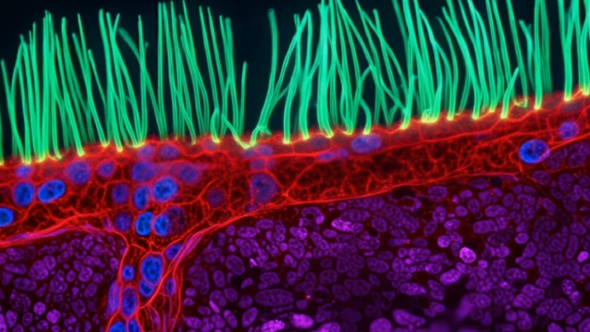
In that sense, the disease is defined by movement, so studying it requires observing those cilia in motion with video, not just still imagery. That’s a challenge, given their small size and rapid movement, plus they must be imaged while still living. That rules out many advanced microscopy techniques, particularly those that rely on intense light or fluorescent dyes.
Xu is an optical engineer whose work involves developing advanced imaging systems for observing cellular structure in motion. He captured the cilia movement using diffractive super-resolution microscopy, which employs multiple lightwaves and a digital micromirror to precisely shape light. This minimizes how much light reaches the living sample and yet can capture the rapid cilia movement at high resolution.
“To understand this disease, you need to observe around 10 seconds of motion, not just a still image,” said Xu. “When people watch cilia beating, they immediately understand that something beautiful and important is happening inside us. I think this competition turns microscopy into a shared language.”
Nguyen Nam Nhat of Vietnam placed second with a video of a tiny roundworm and a single-celled organism; Benedikt Pleyer of Bavaria, Germany, placed third with a video of jellyfish larvae suspended in water droplets; Andrew Moore of Howard Hughes Medical Institute in the US placed fourth with a video of synchronized cell division in neighboring cells; and Patrick Hickey of Hypha Research Ltd. in Edinburgh, Scotland, placed fifth with a video of the dynamics of mitochondria and chloroplasts in Turtle Vine leaf cells.
You can check out those winning entries along with several honorable mentions here.
What's Your Reaction?
 Like
0
Like
0
 Dislike
0
Dislike
0
 Love
0
Love
0
 Funny
0
Funny
0
 Wow
0
Wow
0
 Sad
0
Sad
0
 Angry
0
Angry
0










Comments (0)